
| name | Amanita picea |
| name status | nomen acceptum |
| author | Tulloss, Ovrebo & Halling |
| english name | "Pitch Black Amanita" |
| images |
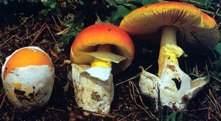
 1. Amanita picea, Andean Colombia. |
| intro |
Amanita picea is a species of the oak forests of Andean Colombia. It is rarely collected; only two collections are known to me. |
| cap |
Its brownish black cap is 23 - 105 mm wide. |
| gills |
The gills of this species are free to very narrowly adnate, close to crowded, and white. The short gills are truncate. |
| stem | It has a 40 - 100 × 10 - 30 mm stem with a pallid ground color that is sometimes decorated with dark fibrils. There is a persistent, skirt-like annulus on the stem. |
| odor/taste | The odor and taste of this species were not recorded. |
| spores |
The spores of this species measure (7.5-) 8.8 - 11.2 (-12.5) × (5.5-) 6.5 - 8.2 (-9.0) µm and are amyloid and broadly ellipsoid to ellipsoid (infrequently subglobose or elongate). No clamps are present at the bases of basidia. |
| discussion |
The stipe's bulbous base is often decorated with rings of volval warts strongly suggesting the arrangement of warts on the lower stipe of Amanita muscaria (L.:Fr.) Lam. The arrangement of the volva and the truncate lamellulae might suggest this species is to be placed in Amanita section Amanita; however, its spores are distinctly amyloid.—R. E. Tulloss |
| brief editors | RET |
| name | Amanita picea | ||||||||
| author | Tulloss, Ovrebo & Halling. 1992. Mem. New York Bot. Gard. 66: 35, figs. 27-28, 36. | ||||||||
| name status | nomen acceptum | ||||||||
| english name | "Pitch Black Amanita" | ||||||||
| MycoBank nos. | 359396 | ||||||||
| GenBank nos. |
Due to delays in data processing at GenBank, some accession numbers may lead to unreleased (pending) pages.
These pages will eventually be made live, so try again later.
| ||||||||
| holotypes | COL; isotype, NY | ||||||||
| intro |
The following text may make multiple use of each data field. The field may contain magenta text presenting data from a type study and/or revision of other original material cited in the protolog of the present taxon. Macroscopic descriptions in magenta are a combination of data from the protolog and additional observations made on the exiccata during revision of the cited original material. The same field may also contain black text, which is data from a revision of the present taxon (including non-type material and/or material not cited in the protolog). Paragraphs of black text will be labeled if further subdivision of this text is appropriate. Olive text indicates a specimen that has not been thoroughly examined (for example, for microscopic details) and marks other places in the text where data is missing or uncertain. The following material not directly from the protolog of the present taxon and not cited as the work of Dr. Z. L. Yang or another researcher is based on original research by R. E. Tulloss. Amanita picea is a medium-sized mushroom. | ||||||||
| pileus | 23 - 105 mm wide, black at first then dark brown (almost black) at center to 6E7 or paler toward margin, becoming shiny when dry, ovoid to hemispheric at first then obtusely convex expanding to plano-convex, becoming virgate in age; context white 4± mm thick, unchanging when cut; margin not striate, nonappendiculate, incurved to decurved; universal veil absent or in detersile, appressed submembranous, fibrillose to cottony, gray to dark brownish gray patches with pallid undersides and margins or in detersile warts, dark brownish gray or (pinkish) gray with darker gray centers and with pallid margins, in all cases developing rusty or fulvous spots. | ||||||||
| lamellae | free to very narrowly adnate, close to crowded, white, becoming pale orangish cream or darker (5A3 to 5A5 or 7.5YR 7-8/6) when dry, 5± mm broad, with margin minutely fimbriate (10× lens); lamellulae truncate. | ||||||||
| stipe | 40 - 100 × 10 - 30 mm, pallid with dense layer of longitudinally arranged gray fibrils, white when young, developing pinkish orange stains with age and near base with handling or as dark as 5C-D4 at maturity, subcylindrical to subventricose; bulb subradicating, splitting longitudinally at broadest point; context white; partial veil subapical to superior, membranous, pendent, flaring, persistent, white above, pale gray below, sometimes striate above; universal veil gray, forming two to three concentric rings (around top of bulb) of coarse floccose minutely verruculose pyramidal to subpyramidal warts with dark peaks, with patch of submembranous gray material (limbus internus?) found on lower stipe of one specimen. | ||||||||
| pileipellis | 225 - 290 µm thick, some brown pigment dissolving in mount (wetted with ethanol, mounted in 2% KOH), extensively gelatinizing; filamentous, undifferentiated hyphae 1.8 - 6.0 µm wide, interwoven, branching, disordered on surface, below surface subradially arranged, many with dark brown intracellular pigment scattered throughout except for hyphae nearest surface and nearest transition to pileus context; vascular hyphae 1.0 - 6.0 µm wide. | ||||||||
| pileus context | filamentous, undifferentiated hyphae 3.2 - 21.5 µm wide, branching, tangled, occasionally coiling, loosely interwoven in open lattice; acrophysalides clavate to elongate to ellipsoid, up to 138 × 80 µm, terminal or in chains (then not so fully inflated), with walls thin or very slightly thickened; vascular hyphae 5.8 - 12.0 µm wide, relatively common. | ||||||||
| lamella trama | bilateral, divergent; wcs = 45 - 65 µm; central stratum including narrowly subfusiform intercalary cells; subhymenial base comprising divergent inflated cells singly or in short chains, diverging in smooth curve, often elongate and apparently all intercalary, mixed with smaller cells of other forms and filamentous, undifferentiated hyphae, with angle of divergence 45° - 60°; filamentous, undifferentiated hyphae 2.0 - 8.5 µm wide, branching; inflated cells ellipsoid to ovoid to elongate, up to 74 × 35 µm, thin-walled; vascular hyphae 1.5 - 4.5 µm wide. | ||||||||
| subhymenium | wst-near = 20 - 50 µm; wst-far = 35 - 65 µm; cellular (pseudoparenchymatous), 5 - 7 cells deep; basidia arising from small ovoid to subglobose to clavate cells in branching chains or from short inflated or uninflated hyphal segments. | ||||||||
| basidia | 37 - 55 × 9.0 - 12.2 µm, 4- or infrequently 2-sterigmate, thin-walled; clamps not observed. | ||||||||
| universal veil | On pileus: filamentous, undifferentiated hyphae 1.0 - 14.0 µm wide, branching, plentiful in upper surface of warts; inflated cells dominant throughout warts except at upper surface, gelatinizing at interface with pileipellis, hyaline or with pale grayish brown intracellular pigment, elongate-ventricose to ellipsoid to ovoid to subglobose, up to 56 × 47 µm, thin-walled, terminal, single or in short chains; vascular hyphae 2.2 - 12.0 µm wide, branching, locally plentiful (especially near upper surface). On stipe base: Very similar to that on pileus, but differing as follows: somewhat more gelatinized and with somewhat larger proportion of filamentous hyphae more commonly in fascicles; inflated cells somewhat larger, up to 73 × 53 µm; vascular hyphae 1.5± µm wide, uncommon. | ||||||||
| stipe context | longitudinally acrophysalidic; filamentous, undifferentiated hyphae 1.8 - 11.5 µm wide, branching, plentiful, often in fascicles of those of smaller diameter; acrophysalides up to 271 × 125 µm, most narrower, with walls thin or very slightly thickened; vascular hyphae 1.5 - 6.5 µm wide, sometimes appearing intermixed in fascicles of filamentous, undifferentiated hyphae, occasionally forming fascicles nearly entirely of vascular elements. | ||||||||
| partial veil | almost entirely comprising interwoven branching filamentous, undifferentiated hyphae 2.5 - 9.5 µm wide, in large part subradially arranged, sometimes in fascicles; inflated cells scarce, terminal, clavate to broadly clavate, up to 59 × 37 µm; vascular hyphae 2.0 - 11.0 µm wide, locally plentiful in knots and tangles. | ||||||||
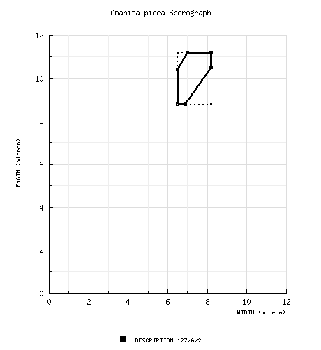
| basidiospores | [127/6/2] (7.5-) 8.8 - 11.2 (-12.5) × (5.5-) 6.5 - 8.2 ( 9) µm, (L = 9.3 - 10.6 µm; L’ = 10.1 µm; W = 6.9 - 7.5 µm; W’ = 7.2 µm; Q = (1.11-) 1.28 - 1.60 (-1.74); Q = 1.35 - 1.47; Q’ = 1.40), amyloid or slightly so, hyaline, colorless, with walls thin or sometimes thickened in apical region, smooth, subglobose to broadly ellipsoid to ellipsoid, occasionally elongate, adaxially flattened or slightly so; apiculus sublateral, cylindric, rather small; contents guttulate; color in deposit unknown. | ||||||||
| ecology | Gregarious to subgregarious. At up to 2700 m elev. Under Q. humboldtii in loamy soil. | ||||||||
| material examined | COLOMBIA: BOYACÁ—Mpio. San Miguel de Sema - rd. from Simijacta to San Miguel de Sema, 9.v.1987 R. E. Halling 5250 (holotype, CO; istoype, NY). NARIÑO—Mpio. Pasto - vereda “La Josefina,” km 17, rd. from Pasto to Chachagüí, 20.xi.1988 A. E. Franco-M. 164 (paratypes; NY & PSO). | ||||||||
| discussion |
In the field, this unique and easily recognizable entity looks like a black form of Amanita muscaria, but it can be separated easily from section Amanita by its amyloid spores. Recent lists of Amanita species known from South America are available (e.g., Raithelhuber, 1986; Garrido & Bresinsky, 1985; Garrido, 1987). In these sources we find no taxon reported in section Validae with a spore length/breadth ratio similar to that computed for A. picea. The world literature contains descriptions of only four taxa in this section with similar spore size, although the values of Q determined for A. picea are very commonly found in species of section Validae. The four taxa with similar spores are macroscopically quite dissimilar from A. picea:
Amanita morrisii Peck of eastern North America is somewhat similar to A. picea in the colors of its pileus in early stages of expansion. Tulloss has seen unpublished watercolors of topotypes made by the original collector (G. E. Morris) and now located in PM and NYS; these paintings show a pinkish underside to the annulus, a nearly naked bulb, and very pale submembranous (not wart-like) patches of universal veil material. Tulloss (1991) reported the spores of A. morrisii to be [485/24/6] (6-) 7.2 - 9.5 ( 11.5) × (4.2 ) 5.5 - 7 (-8.2) with Q = (1.28-) 1.30 - 1.39 (--1.42). | ||||||||
| citations | —R. E. Tulloss | ||||||||
| editors | RET | ||||||||
Information to support the viewer in reading the content of "technical" tabs can be found here.
Each spore data set is intended to comprise a set of measurements from a single specimen made by a single observer; and explanations prepared for this site talk about specimen-observer pairs associated with each data set. Combining more data into a single data set is non-optimal because it obscures observer differences (which may be valuable for instructional purposes, for example) and may obscure instances in which a single collection inadvertently contains a mixture of taxa.

Text and User-Generated Sporographs are published under the Creative Commons License.

In the case of a taxon page, image credits are on the 'image' tab.